New products and Services
Two great non-mydriatic fundus cameras new to Grafton
Two great non-mydriatic fundus cameras new to Grafton
Fundus Vue from CrystalVue
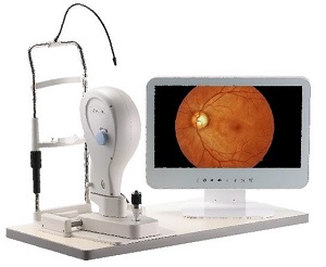 Grafton Optical announce the launch of the FundusVue from CrystalVue, providing vivid color, high-definition retinal images as an essential diagnostic aid to clinicians.
The latest generation of non-mydriatic fundus camera is designed to capture and provide images of the eye as an aid to clinicians in the diagnosis of diabetic retinopathy*, AMD, glaucoma and other retinal diseases. This modern ophthalmic instrument combines a simple method of image capture with superior image quality in a fast and efficient manner. It is easy to operate, and is ultra compact and portable in terms of design.
Fast image capture
The dedicated FundusVue software speeds up image capture with live preview imaging. The entire process, from welcoming the patient to delivering high quality images, takes only a few minutes.
Easy to use
Multiple controls and settings are not necessary with the FundusVue. Internal fixation lights and self-guiding software assist the user in capturing high quality images. Simply align the pupil, optimise the focus in the live on screen preview and capture the image. Exposure and LED flash are automatically controlled for most patients.
Portable
Weighing only 2Kg, FundusVue’s compact design saves on setup space and can be easily transported wherever needed. The simple USB connection allows flexibility as to where examinations take place. It also makes it easy for the user to review the images with their patient.
Retinal imaging software
The bundled software allows users to store, retrieve, archive, process and share the digital images instantly, with just a mouse click. Images can also be exported to the storage server and output to a printer. Features include: colour, digital red-free, RGB and cup-to-disc.
* FundusVue is not currently on the list of approved devices for the NHS National Diabetic Eye Screening Program.
NFC-600 from CrystalVue
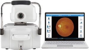
NFC-600 from CrystalVue is a portable, fully automatic, all in one non-mydriatic retinal camera; providing high quality retinal images for remote screening areas and telehealth.
With automatic 3D tracking and focusing, retinal images can be captured with a single tap. The full-auto-shot function shortens examination times, which not only simplifies the examination process, but also reduces discomfort or strain for patients. The NFC600 is an essential diagnostic instrument for practitioners to evaluate and record a patient’s retina.
High quality retinal images
With high resolution of 12 million pixels, the NFC-600 captures and generates high quality retinal images. It provides retinal diagnostic staff and AI systems with more precise and helpful information, which increases diagnostic accuracy and efficiency. The image can be enlarged to see tiny details. Users can also change colours or apply photo effects to the image for different purposes. Ten internal fixation targets are selectable. The disc, fovea, macular, or other peripheral retina areas can be captured by selecting the specified fixation.
Enhanced portability
With its compact design, the NFC-600 can be easily moved around and set up for use in different locations. The simple USB interface offers flexible connectivity to operate the retinal camera. The head and body of the device are designed to be detachable for better portability and an optional customised wheeled moving case is also available for the NFC-600, which increases mobility.
Enhanced connectivity
NFC-600 can be easily connected to any Windows-based PC or laptop with simple USB connection. It is also DICOM compliant, making it easy to integrate with PACS program.
Built-in filters
NFC-600 provides red-free, negative film, RGB, grey scale filters for users to change or apply photo effects to the image for different purposes.

